Pulsating Exophthalmos
🔍 Definition
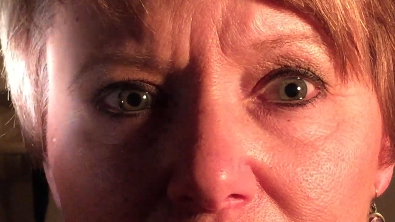
Pulsating exophthalmos is the rhythmic forward movement of the eyeball that occurs synchronously with the pulse, often accompanied by a bruit (a whooshing sound) heard over the orbit.
🧠 Causes (Major Etiologies)
1. Carotid-Cavernous Fistula (CCF) – Most common cause
Abnormal communication between carotid artery and cavernous sinus.
Causes arterial blood to flow into the venous system → orbital venous congestion → pulsation.
Often traumatic or spontaneous (aneurysmal rupture).
2. Orbital Arteriovenous Malformation (AVM)
Congenital AV shunt in the orbit causing increased flow and pulsation.
3. Meningocele / Meningoencephalocele
Herniation of meninges or brain tissue through a defect in orbital wall → transmitted pulsations from intracranial contents.
4. Neurofibromatosis (Type I)
Congenital defect of the sphenoid bone → pulsation transmitted directly from intracranial cavity.
⚕️ Clinical Features
Feature Description
Pulsation Synchronous with the heartbeat
Bruit Audible on auscultation over orbit; may disappear with carotid compression
Proptosis Usually reducible by gentle pressure or carotid compression
Conjunctival changes Chemosis (swelling), dilated tortuous veins, redness
Ocular movements Restricted due to congestion
Vision May be decreased due to optic nerve compression or retinal venous stasis
Pain Common in traumatic cases (e.g., CCF)
🧩 Differential Diagnosis
Condition Key Feature
Simple Exophthalmos Non-pulsatile (e.g., thyroid eye disease)
Thrill Exophthalmos May have palpable thrill in CCF
Intermittent Exophthalmos Appears during venous congestion or Valsalva (e.g., orbital varix)
🩻 Investigations
CT / MRI Orbit & Brain – Evaluate bone defects, vascular malformations, soft tissue changes.
CT/MR Angiography – Identify CCF or AVM.
Carotid Angiography (Digital Subtraction Angiography) – Gold standard for diagnosing carotid-cavernous fistula.
Ophthalmoscopy – Dilated veins, optic disc edema.
💊 Treatment
Depends on the cause:
Carotid-Cavernous Fistula
Endovascular embolization (preferred)
Ligation of carotid artery (rarely used now)
Orbital AVM
Embolization or surgical excision
Sphenoid Bone Defect (Neurofibromatosis)
Surgical repair if progressive
Supportive Care
Lubricating drops
Control of intraocular pressure
Manage exposure keratitis
🧬 Mnemonic for Causes
“CAMe N”
C – Carotid-cavernous fistula
A – AV malformation
Me – Meningocele / Meningoencephalocele
N – Neurofibromatosis (sphenoid defect)


Medical Student
This was incredibly helpful for my upcoming exam. Thank you!
Nursing Professional
Great explanation of the ECG changes in hyperkalemia!